
A cluster of lab-grown mouse neurons — no body, no senses, no lived experience — just learned how to keep a virtual pole from falling over. And it only took 45 minutes.
Researchers at the University of California, Santa Cruz have trained brain organoids, sometimes called “mini-brains,” to tackle the cart-pole problem, one of the most well-known benchmark challenges in control engineering. The result was a 46% success rate under adaptive feedback training, compared to just 4.5% under random training. That’s a tenfold improvement, coming entirely from living tissue in a dish.
The numbers alone are striking. But what this experiment actually reveals about the nature of learning — and what it could mean for understanding some of the most debilitating neurological conditions humans face — is the part of the story that deserves the most attention.
What the Cart-Pole Problem Actually Tests
The cart-pole problem is a classic in engineering and artificial intelligence research. Imagine a pole balanced upright on a cart that can move left or right. The goal is to keep the pole from tipping over by adjusting the cart’s position in real time. It sounds simple, but it requires continuous feedback, rapid adjustment, and something that looks a lot like learning from mistakes.
It’s the kind of task used to benchmark AI systems precisely because it demands dynamic, adaptive responses — not just pattern recognition. Using it to test biological tissue takes that benchmark into genuinely new territory.
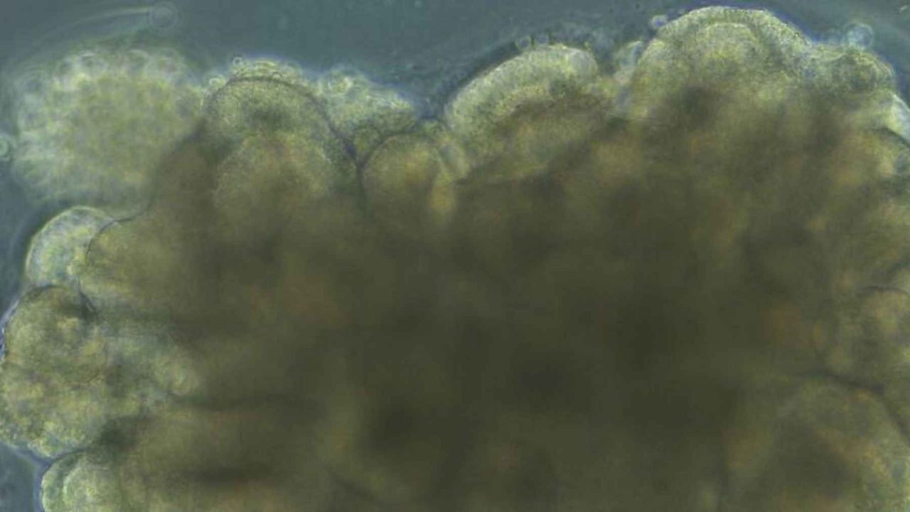
The organoids used in this study were derived from mouse brain cells, grown into small three-dimensional clusters of cortical neurons. They weren’t connected to a robot or a physical cart. Instead, they were given electrical stimulation as feedback signals and allowed to respond. Under adaptive training, where the feedback adjusted based on their performance, the organoids steadily improved over the 45-minute session.
The Real Discovery: What This Tells Us About Living Brain Tissue
The headline result is impressive, but the researchers say the deeper finding is biological, not computational. The study suggests that neural plasticity — the ability of neurons to reorganize and strengthen connections based on activity — may be intrinsic to living cortical tissue itself, even in highly simplified lab models like organoids.
In other words, you don’t need a full brain, a nervous system, or years of sensory experience for neurons to start adapting to a task. The capacity for learning-like behavior appears to be baked into the basic biology of cortical cells at some fundamental level.
That’s a significant finding for neuroscience. It raises questions about where exactly the boundary between “learning” and simple biological responsiveness lies — and whether current definitions of artificial intelligence are measuring the right things at all.
Key Findings at a Glance
| Training Condition | Success Rate | Training Duration |
|---|---|---|
| Adaptive feedback training | 46% | 45 minutes |
| Random training (control) | 4.5% | 45 minutes |
- Organoids were derived from mouse brain cells, grown into cortical tissue clusters
- The task used was the cart-pole problem, a standard engineering benchmark
- Research was conducted at the University of California, Santa Cruz
- The improvement factor between adaptive and random training was roughly tenfold
- Neural plasticity was identified as the likely mechanism behind the performance gains
Why This Matters for Alzheimer’s, Parkinson’s, and Beyond
The practical implications extend well beyond the lab bench. Researchers say the findings could help scientists study how learning is disrupted in conditions including Alzheimer’s disease, Parkinson’s disease, schizophrenia, and ADHD.
Each of those conditions involves, in different ways, a disruption to normal neural plasticity. If organoids can be trained to demonstrate measurable learning behavior, they could become a powerful model for observing exactly how and when that plasticity breaks down — without requiring animal studies or waiting for human clinical data.
That’s a meaningful step forward. Currently, studying learning impairment in neurodegenerative diseases is extraordinarily difficult because researchers are often working backward from symptoms that appear years or decades after the underlying cellular changes begin. A reliable lab model that demonstrates learning at the cellular level could change the timeline for that research significantly.
For patients and families living with these conditions, the path from lab finding to treatment is always long. But the ability to observe neural plasticity failing in real time, in a controlled environment, is the kind of foundational research that eventually makes new treatments possible.
The Question This Raises About Artificial Intelligence
There’s a deeper philosophical thread running through this research. Modern AI systems — including the large language models and reinforcement learning agents that dominate headlines — are built on mathematical models loosely inspired by the brain. But they don’t use living tissue, they don’t have biological plasticity, and they learn through computational processes that are fundamentally different from what neurons do.
When a cluster of lab-grown neurons outperforms a random baseline on a task that AI researchers use as a standard benchmark, it naturally prompts the question: what exactly is learning, and are our machines actually doing it? The organoid study doesn’t answer that question, but it makes it harder to dismiss.
The researchers’ framing is careful — they’re not claiming the mini-brain is conscious, sentient, or truly “thinking.” What they are claiming is that the plasticity observed is real, measurable, and appears to emerge naturally from cortical tissue under the right conditions. That’s a meaningful distinction, and an honest one.
What Comes Next for This Research
The University of California, Santa Cruz study opens several directions for follow-up work. Researchers now have a working framework for using organoids as learning models, which means future experiments could test more complex tasks, longer training windows, or organoids derived from human cells rather than mouse cells.
The disease-modeling applications — particularly for Alzheimer’s and Parkinson’s — are likely to attract significant scientific interest in the near term, given how urgently better research models are needed in those fields.
Whether this line of research eventually contributes to next-generation computing architectures that incorporate biological components remains an open question. For now, the most immediate value is in what it teaches us about the brain we already have.
Frequently Asked Questions
What is a brain organoid?
A brain organoid is a small, three-dimensional cluster of neurons grown in a laboratory from biological cells. In this study, the organoids were derived from mouse brain cells and used as simplified models of cortical tissue.
What was the cart-pole problem used in this study?
The cart-pole problem is a classic engineering benchmark where a virtual pole must be kept upright on a moving cart through continuous real-time adjustments. It is widely used to test adaptive and learning-based systems.
How much better did the organoids perform under adaptive training?
Under adaptive feedback training, the organoids achieved a 46% success rate, compared to just 4.5% under random training — roughly a tenfold improvement over the 45-minute session.
Which diseases could this research help with?
Researchers say the findings could help scientists study how learning is disrupted in Alzheimer’s disease, Parkinson’s disease, schizophrenia, and ADHD, all of which involve disruptions to normal neural plasticity.
Does this mean the mini-brain is conscious or thinking?
The researchers make no such claim. The study focuses specifically on measurable neural plasticity and learning-like behavior in cortical tissue, not on consciousness or sentience.
Where was this research conducted?
The research was conducted at the University of California, Santa Cruz.


Leave a Reply